
April 2, 2026 | Jeanna Vazquez
Transforming High-Quality Care at East Campus Medical CenterThe newest hospital at UC San Diego Health is delivering world-class care through a unique service model led by physicians, nurses and staff.

April 2, 2026 | Jeanna Vazquez
Transforming High-Quality Care at East Campus Medical CenterThe newest hospital at UC San Diego Health is delivering world-class care through a unique service model led by physicians, nurses and staff.

March 26, 2026 | Jeanna Vazquez
Emily Lukacz, MD, Named President of American Urogynecologic SocietyAs a board-certified urogynecologist, Lukacz specializes in diagnosing, treating and studying female pelvic floor disorders, such as incontinence and prolapse.

February 24, 2026 | Leslie Aquinde
New Blood Test Score Detects Hidden Alcohol-Related Liver DiseaseNew blood test score helps detect hidden alcohol-related liver disease early, offering a simple cost-effective tool to guide timely care.
At UC San Diego Health, you'll experience compassionate care that puts you first. From diagnosis to recovery and beyond, our experts are leading the way in innovation and bringing you the latest in treatment options.

Flu season lasts from fall to spring. Learn key symptoms, when to see a health care provider, and get same-day flu testing and treatment at UC San Diego Health Express Care.

Get expert answers to common flu shot questions to help you stay informed, healthy and protected throughout flu season.
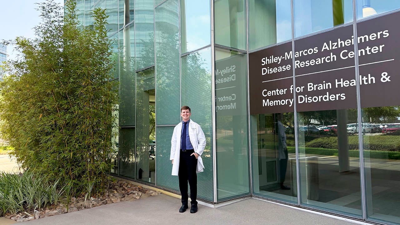
Ian Neel, MD, has been named co-chair of the San Diego Alzheimer’s Project. In this Q&A, he reflects on his journey to cognitive care for older adults.

A story of ALS care, community strength and hope at UC San Diego Health.

This patient story follows Dr. Sunny Sharma's acoustic neuroma diagnosis, surgery, gamma knife treatment and recovery to marathon running.

Discover personalized neurosurgery at UC San Diego Health, treating brain, spine, and peripheral nerve conditions with advanced technology and expertise.

UC San Diego Health patient’s journey through chemotherapy radiation, surgery, a clinical trial for larynx cancer.

Know the early warning signs of colorectal cancer, when to get screened starting at 45, and why early detection can save lives.

First global trial shows tumor DNA-guided cancer therapy is safe and effective, improving outcomes through personalized drug combinations in oncology care.

UC San Diego Health registered dietitian answers commonly asked questions about how to eat right for heart health — including healthy swaps and a recipe.
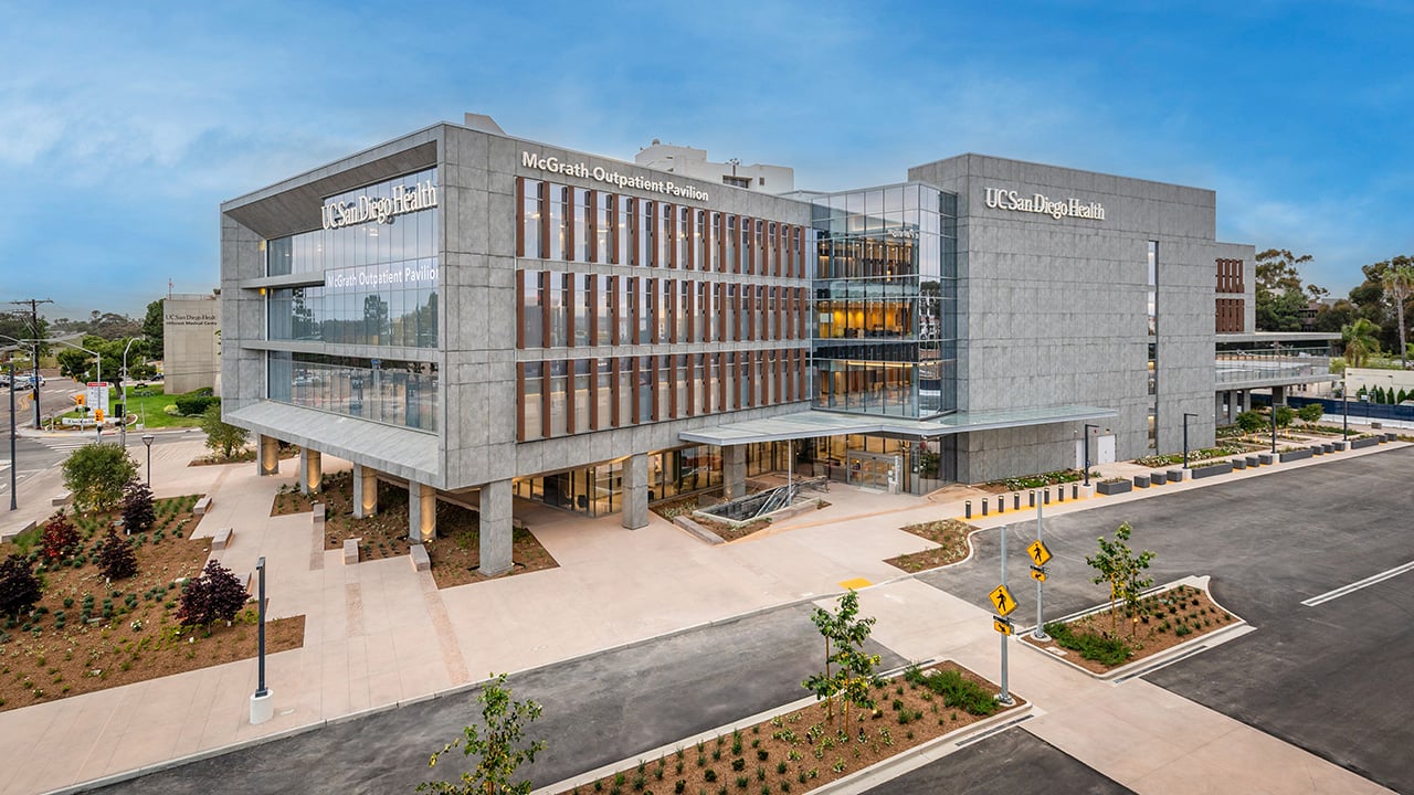
McGrath Outpatient Pavilion opens at UC San Diego Health, expanding access to advanced specialty care, surgery and cancer treatment in Hillcrest.
UC San Diego Health heart specialists treat over 1,000 people each year with pacemakers or ICDs, offering expert care for heart rhythm disorders.
As a board-certified urogynecologist, Lukacz specializes in diagnosing, treating and studying female pelvic floor disorders, such as incontinence and prolapse.

For the fourth year in a row, UC San Diego Health is recognized as a Best Hospital for Maternity Care by U.S. News & World Report.

UC San Diego Health is the first health system in San Diego to receive national accreditation to offer in-clinic moderate sedation for patients.

Joint powers authority will stabilize and expand health care services for the community in the Palomar Health District of North San Diego.

UC San Diego Health recognized by Vizient as a top performer in hospital quality and outpatient care for the seventh year.

Behind the scenes with Lisa Rhodes as she shares what it took to bring McGrath Outpatient Pavilion to life and what it means for the future of health care.