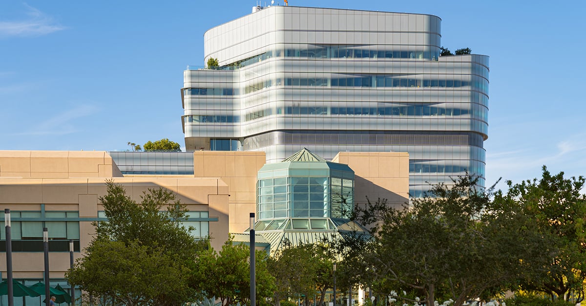
Researchers at the University of California, San Diego (UCSD) are conducting cutting-edge research in Magnetic Resonance Imaging (MRI) that could impact patient care here and around the world in coming years. A new facility to house this research effort, the 3Tesla Magnetic Resonance (MR) Research Laboratory at the UCSD Medical Center, Hillcrest, will celebrate its grand opening on May 2.
The centerpiece of the research space is the new General Electric Signa 3Tesla whole body magnet with a magnetic field strength of 3Tesla, approximately 60,000 times stronger than the earth’s magnetic field.
“This state-of-the-art imaging machine will enable us to visualize tissue that has been previously undetectable by MR imaging,” said Graeme Bydder, M.B., Ch.B., UCSD professor of radiology and director of the new laboratory. Bydder is one of the world’s pioneers in the development of MRI for clinical use in patients – a technology now almost taken for granted, but arguably the greatest advance in diagnostic medical techniques over the past century.
MRI is a painless and safe diagnostic procedure that uses a powerful magnet and radio waves to produce detailed images of the body's organs and structures, without the use of X-rays or other radiation. Signals from the MRI scan are converted by computer into a series of extremely clear, cross-sectional images. The computer is able to reconstruct all the images into a single picture or into three-dimensional images, allowing complete visualization of the body area scanned from all angles. However, until recently, most clinical MRI has only been able to image soft tissues such as liver, lungs and brain. It had not been possible to get images of semi-solid tissue such as ligaments, tendons, cartilage or the fibrous membrane on the surface of bones.
Under Bydder’s leadership, the UCSD team of radiologists is improving the MR techniques for viewing the musculoskeletal system, as well as the brain, liver and breast, which will enable clinicians to better diagnose and treat injury to ligaments and tendons and diseases including breast and liver cancer, fatty liver disease and cirrhosis.
“Work by our researchers is changing the field of MRI by developing techniques that will allow clinicians to view never-before-seen images of soft tissue, with today’s technology detecting signals 100 to 1,000 times earlier than previously possible with clinical MR systems,” said William G. Bradley, Jr., M.D., Ph.D., F.A.C.R., professor and chair of radiology at UCSD’s School of Medicine.
In addition, among Bydder’s current research projects is the clinical application of a technique he developed to obtain images of previously MR-invisible, semi-solid tissues such as cortical bone, ligaments and tendons. These tissues appeared as “black holes” with traditional MR Imaging. His new technique allows physicians to see solid tissue and different components within them.
Christine B. Chung, M.D., associate professor of musculoskeletal radiology, is conducting cutting-edge imaging of soft tissues, allowing clinicians to evaluate damage to bones and joints as never before. New imaging techniques developed by Chung and other UCSD researchers will enable clinicians to clearly view the deep layers of cartilage, enabling them to detect injuries earlier and with greater detail, thus avoiding development of degeneration to these tissues. Such imaging also helps diagnosis of rheumatologic disorders such as arthritis, giving physicians the ability to detect inflammation present in early degeneration and to monitor therapy to see how it impacts damage to soft tissue.
Claude Sirlin, M.D., an assistant professor of radiology, is chief of Body MRI, head of the Liver Imaging Research Group and head of clinical research in the Division of Body Imaging at UCSD. The Liver Imaging Research Group was formed in 2004 to develop new techniques for liver imaging. Among his goals are to optimize clinical MR imaging protocols for hepatocellular carcinoma, the most common form of liver tumor; improved detection of liver cirrhosis, and improved assessment of patients with fatty liver disease.
Michael Middleton, M.D., Ph.D., assistant clinical professor of radiology, has more than 15 years experience in breast MR imaging first with breast implant imaging and more recently, with breast cancer. UCSD has performed nearly 2,000 breast MR scans with breast cancers or suspected cancers, more than any other hospital system in San Diego. Middleton is looking forward to conducting research in breast cancer at the new lab using the 3T magnet, which gives physicians the opportunity to see tumors much earlier and better characterize them.
“This is very important in light of recommendations at the international level on how and when breast MR is recommended,” said Middleton. “An additional one million MR studies per year are anticipated in the United States alone in the coming year, with demand greatly outweighing supply of imaging resources,” he said, adding that physicians have not yet seen the peak of the demand curve for breast MR imaging.
# # #
Media Contact: Debra Kain, ddkain@ucsd.edu or Kimberly Edwards, edwards@ucsd.edu, 619-543-6163