Brain Imaging
If your doctor recommended a brain imaging scan at UC San Diego Health, this page explains what it is, how it works and how to prepare.
What Is Brain Imaging?
Brain Imaging involves using advanced imaging techniques, such as MRI, CT and PET scans, to evaluate the structure and function of the brain.
These techniques are crucial for diagnosing neurological conditions and monitoring treatment progress.
How It Works
MRI
This test uses powerful magnets and radio waves to produce detailed images of the brain, especially helpful for viewing soft tissues.
CT
This imaging technique uses X-rays to create cross-sectional images of the brain. CT scans are commonly used to detect bleeding, tumors and structural abnormalities.
PET
A positron emission tomography (PET) scan involves injecting a small amount of radioactive tracer to visualize and evaluate how the brain is functioning, including its metabolism and blood flow.
Common Uses of Brain Imaging
- Diagnosing brain tumors and other abnormalities
- Detecting and evaluating strokes
- Assessing brain injuries and trauma
- Monitoring neurological conditions such as Alzheimer's disease or multiple sclerosis
Preparing for Your Brain Imaging
- Clothing: Wear comfortable clothes without metal snaps, zippers or buttons. If your clothing contains metal, you may be asked to change into a gown.
- Remove metal: Take off jewelry, watches, eyeglasses and other metal objects before the scan.
- Medical devices: Let the imaging team know if you have any implanted devices, such as a pacemaker. Special instructions may be needed.
- Fasting: If you're having a PET scan, you may need to fast for four to six hours beforehand.
After Your Brain Imaging
A radiologist will analyze and interpret the images, and your doctor will go over the findings with you. Depending on the results, you may need additional imaging or treatment.
Patient Help Center
We know that navigating health care can feel overwhelming. Our team is here to make your experience as smooth and supportive as possible. Below, you’ll find resources to guide you every step of the way, from scheduling your appointments to accessing your results.
Advanced Imaging, Trusted Results
At UC San Diego Health, our specialized expertise, advanced technology and collaborative approach ensure fast, accurate imaging services. Our team-based model empowers you and your doctors with the precise information needed to make confident, informed treatment decisions. Explore our imaging services:
Driving Innovation to Transform Lives
UC San Diego Health is at the forefront of advancing imaging technology to enhance patient care.
Our breakthroughs are reshaping health care — making it more efficient, accessible and personalized — so that every patient receives the highest quality of care.

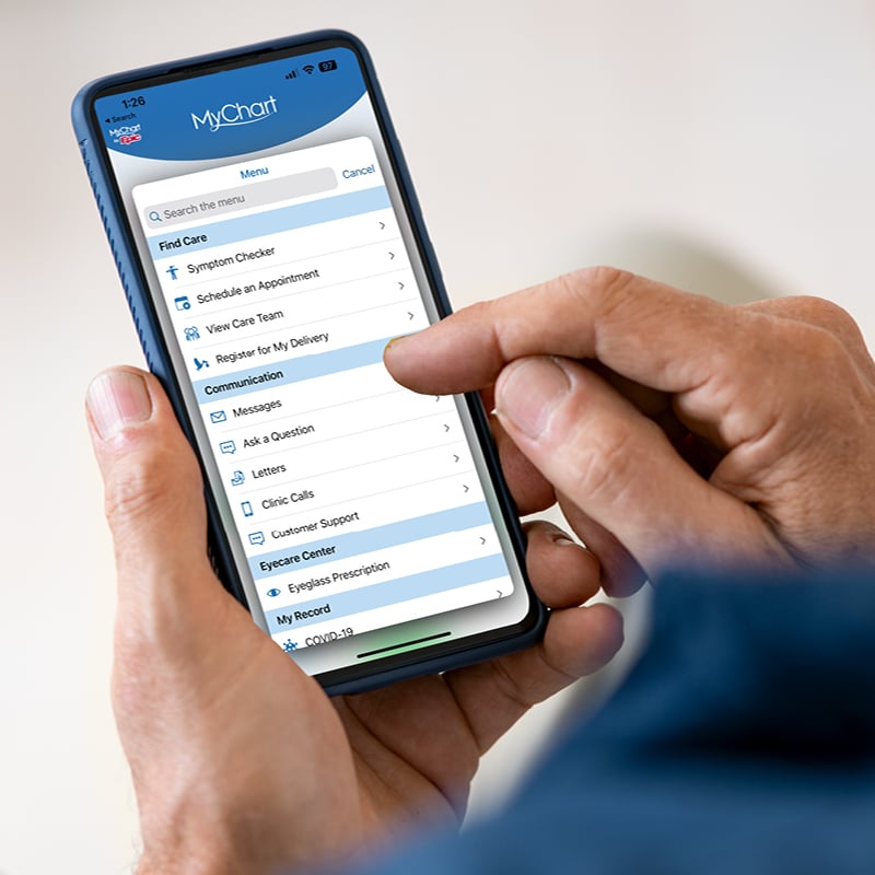
Download MyUCSDHealth App
Use our free app to access records on your phone or tablet.
Download MyUCSDHealth App
Use our free app to access records on your phone or tablet.